Product Description
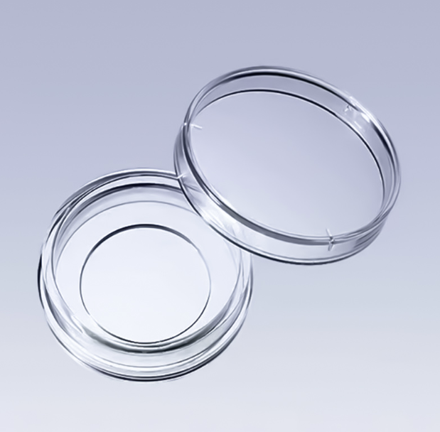
The cell culture dish is made of pure medical grade polystyrene material, which is subject to strict process control and ergonomic design to maximize the safety, efficiency and convenience of cell culture experiment to meet the needs of all users.
AI Product Description
*The following content is generated by AI and is for reference only.
The product described is a specialized glass-bottom cell culture dish designed for advanced microscopy applications. Unlike standard polystyrene dishes, these vessels feature a high-quality, ultra-thin borosilicate or quartz glass bottom, which provides exceptional optical clarity and minimal light refraction. This unique construction makes them indispensable for researchers conducting high-resolution imaging techniques such as confocal laser scanning microscopy (CLSM), total internal reflection fluorescence (TIRF), super-resolution microscopy, and live-cell time-lapse studies. The thin glass substrate allows deep penetration of excitation light and efficient collection of emitted signals, ensuring that fine cellular structures, organelles, and dynamic processes are captured with maximum fidelity.
These dishes typically come in various sizes to accommodate different experimental scales while maintaining compatibility with standard incubators and microscope stages. They are often treated with specific surface coatings, such as poly-L-lysine, collagen, or fibronectin, to enhance cell adhesion and spreading, particularly for sensitive primary cells or neurons that require a stable environment for growth. The use of glass bottoms eliminates the spherical aberration often caused by thick plastic walls, thereby improving image quality at high magnifications.
When handling these delicate instruments, strict adherence to safety and procedural guidelines is essential. First, always inspect the glass bottom for micro-cracks or scratches before use, as any imperfection can compromise both the structural integrity and the optical performance during observation. Handle the dishes by the rim only to avoid fingerprints or oils on the viewing area, which can scatter light and degrade image resolution. During washing or sterilization, never use abrasive materials; instead, rely on validated autoclaving protocols if reusability is intended, though many are supplied pre-sterilized via gamma irradiation for single-use convenience. Furthermore, ensure that the media volume does not overflow the edges to prevent contamination or damage to the microscope objective lens. Proper storage in a dust-free environment is also critical to maintain the pristine condition of the optical surface. By following these protocols, scientists can maximize the utility of these premium tools for breakthrough discoveries in cell biology and biomedical research.





 (All Rights Reserved)